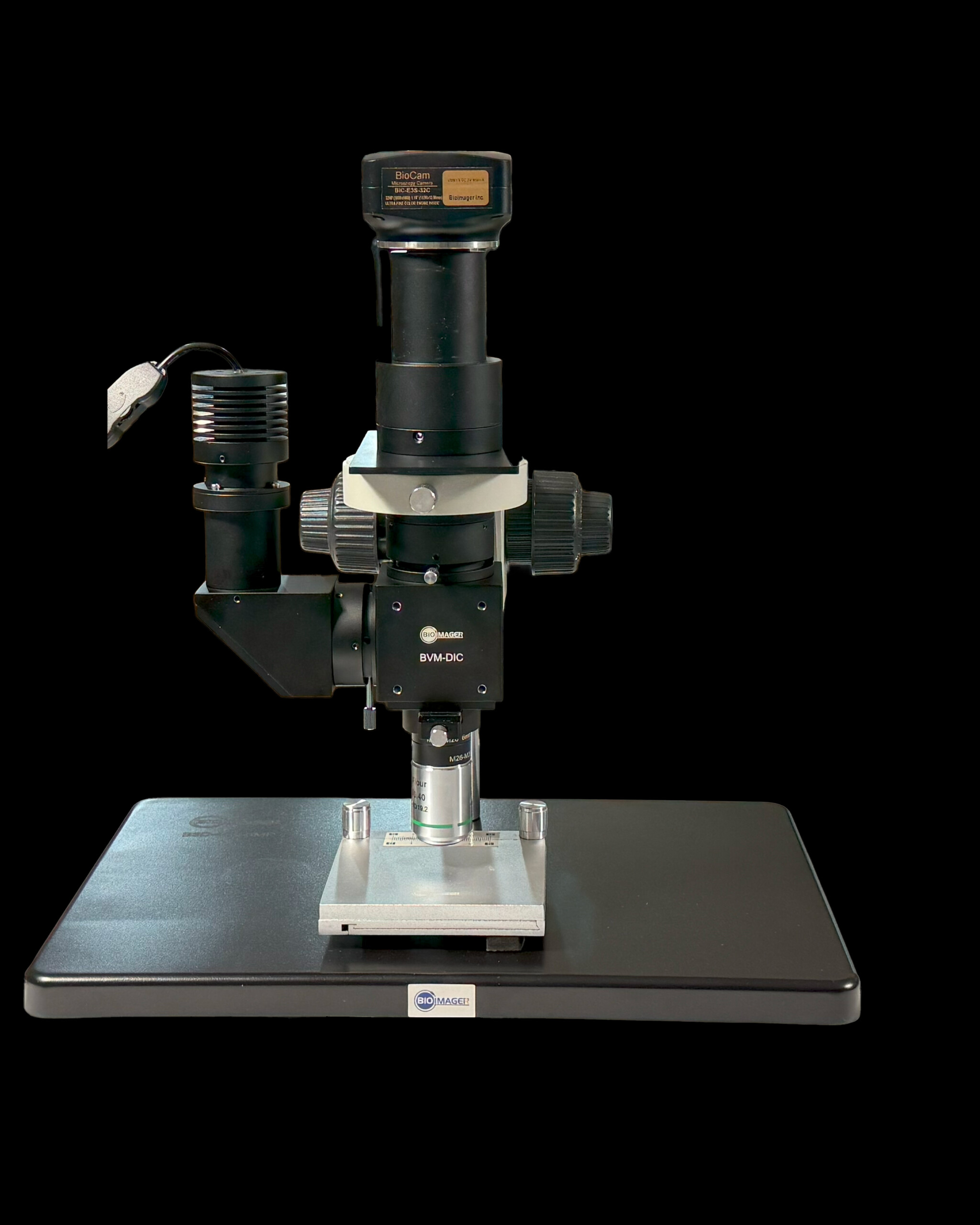
DIC Nomarski Video Inspection Microscope, BVM-DIC Series
Explore the advanced BVM-DIC Microscopy System for high-resolution imaging, enhanced contrast, and precise material analysis. Upgrade your research capabilities today.
The BVM-DIC Series Differential Interference Contrast Microscopy System is an essential tool for advanced research. Utilizing dual-beam polarization interference, it delivers high-resolution imaging with enhanced contrast, ideal for detecting conductive particles, surface cracks, and microbial cells. The system’s adjustable Nomarski prism allows for optimal brightness and interference color adjustments, providing superior observation.
Key features of the BVM-DIC include magnification options up to 50X, multiple objective lenses, and versatile lighting choices, such as 10W white or blue LEDs. This system excels in precise material analysis, ensuring accurate detection of fine structures, defects, and living cell activity.
Whether for conductive particle detection in LCD/OLED circuits or observing microbial cell activity, the BVM-DIC Series offers unparalleled detail and clarity. Its high-resolution capabilities clearly display intracellular contours and structures, making it perfect for various research applications. Upgrade your microscopy research with the BVM-DIC Series and experience the pinnacle of imaging performance.
Features
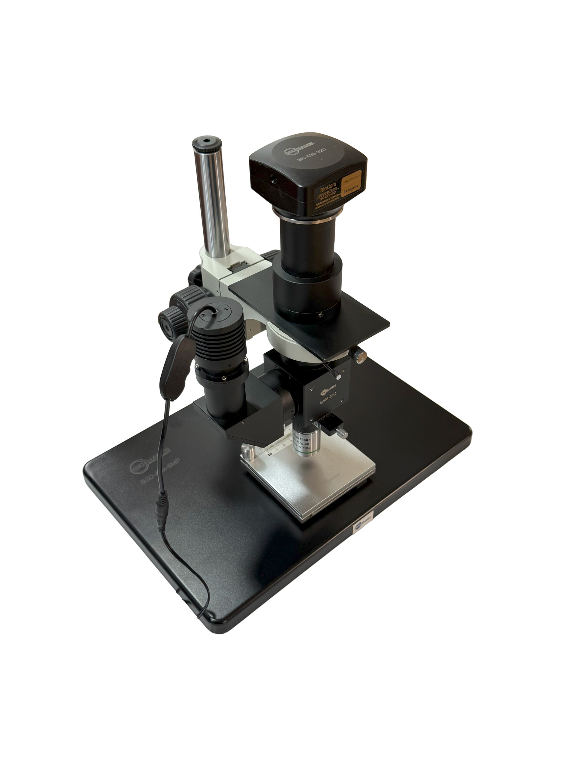
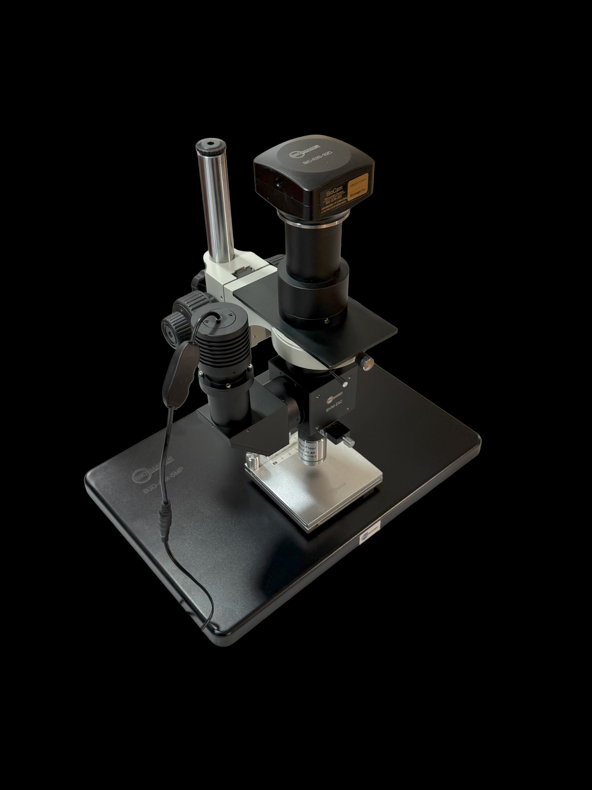
Portable DIC Nomarski Imaging Microscope:
World’s Smallest DIC Module: Compact and highly portable, setting a new standard in microscopy technology.
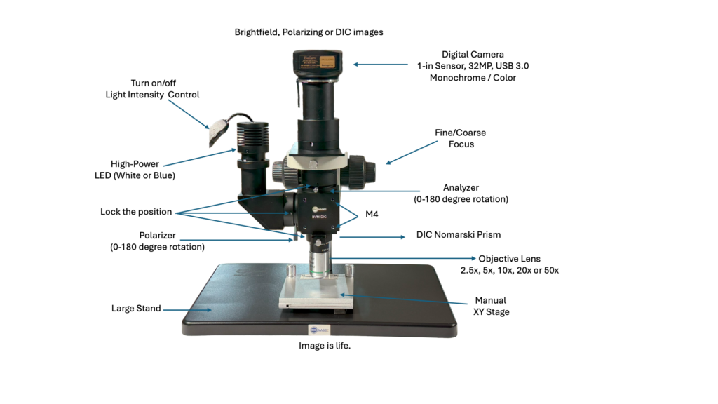
Versatile Objective Lenses: Includes 2.5x, 5x, 10x, 20x, or 50x DIC objective lenses for detailed imaging across multiple scales.
Multiple Imaging Modes: Equipped with capabilities for Brightfield, polarization, and DIC (Differential Interference Contrast) imaging modes, enhancing visual clarity and contrast.
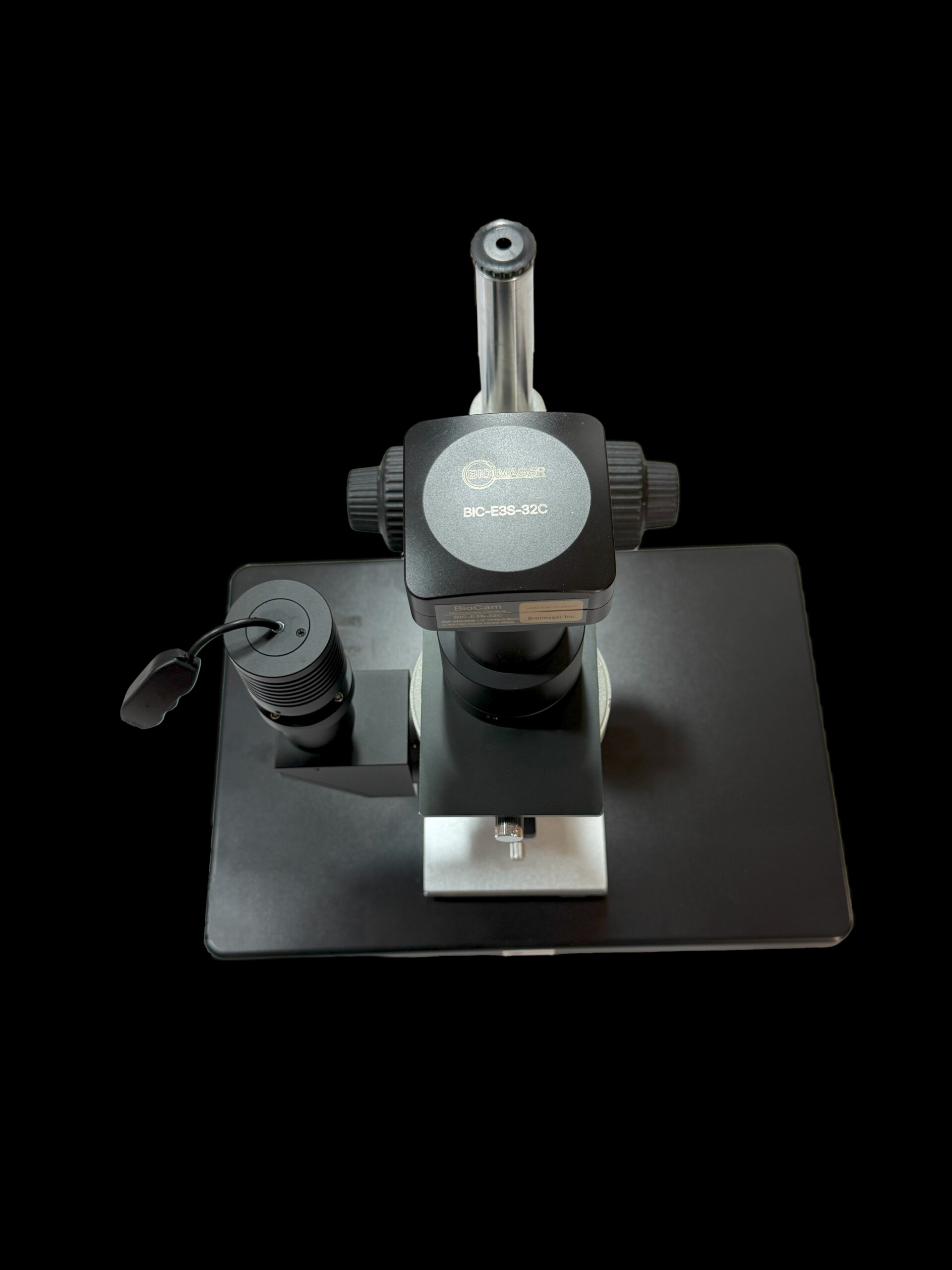
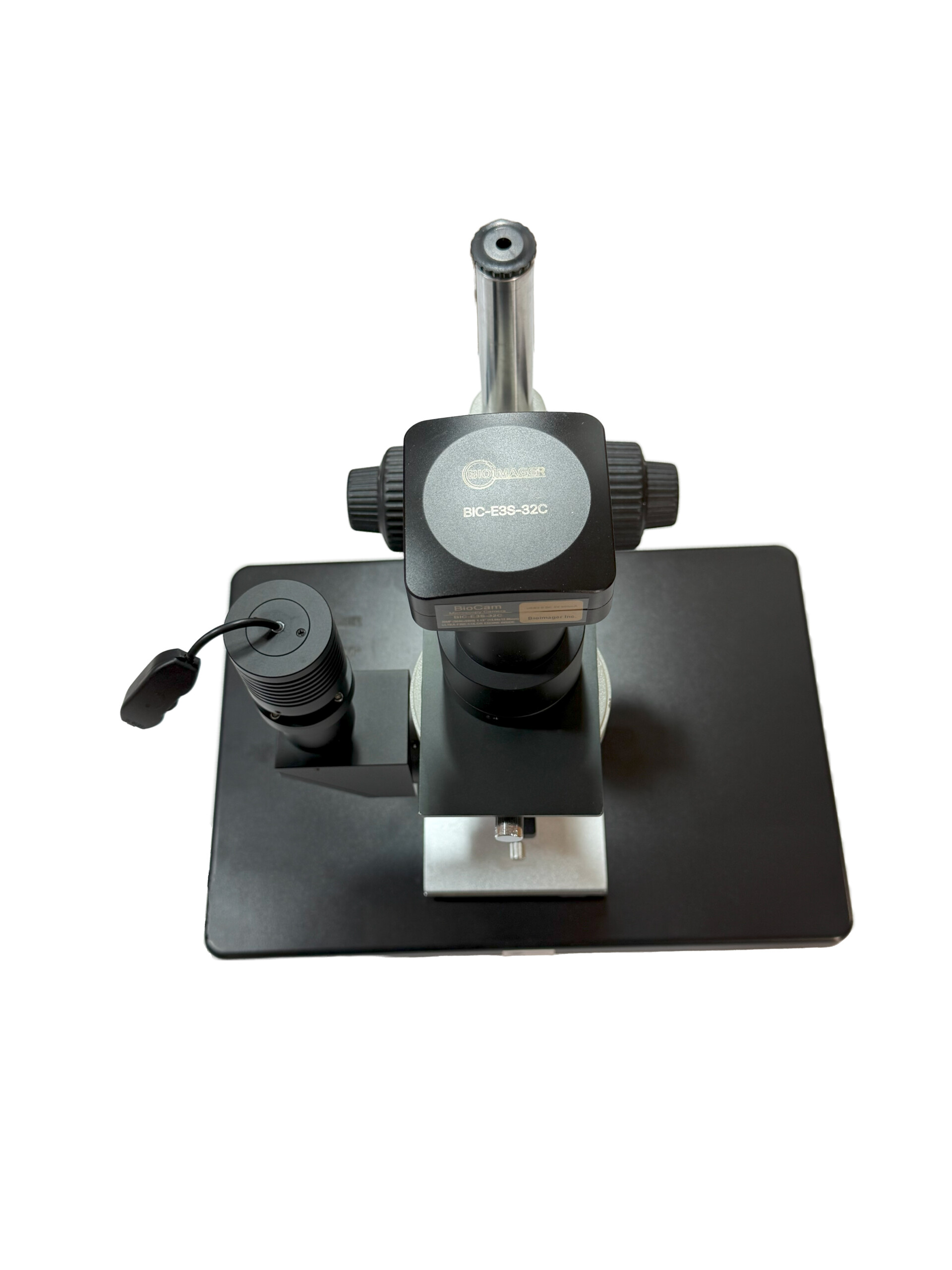
Camera Compatibility: Features connectivity for a 1-inch sensor camera, facilitating high-quality image capture.
Advanced Focusing System: Comes with an optional fine/coarse focus mechanism, allowing for precise adjustments and stable imaging.
Adjustable Optical Components: Offers zero to 180-degree rotations of the polarizer and analyzer with an easy lock mechanism for the polarizer, analyzer, and DIC prism positions, ensuring optimal alignment and stability.
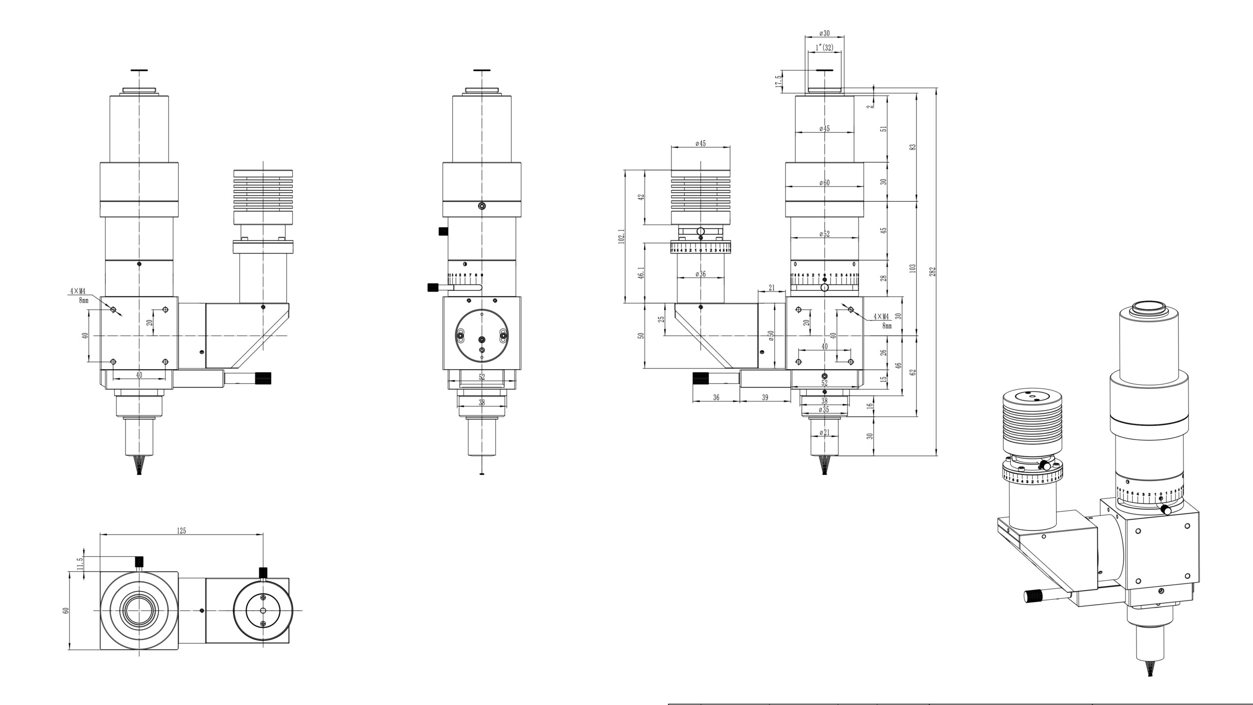
Easy Integration: Includes four M4 threaded holes spaced 40mm x 40mm apart for straightforward mounting on machinery, enhancing versatility in application settings.
Customization Options:
Available customization for inverted mode, making it suitable for a wide range of microscopy applications.
Mini or Macro XY stage
Small or large Stand
This portable microscope combines flexibility with advanced features, making it ideal for both field and lab applications.
The BVM-DIC Series Differential Interference Contrast (DIC) Microscopy System is designed for high-precision optical imaging, offering exceptional contrast enhancement and detailed visualization of microscopic samples. Utilizing dual-beam polarization interference, this Nomarski DIC microscope provides 3D-like imaging with superior resolution, making it an essential tool for advanced microscopy applications.
How the BVM-DIC Microscope Works?
Polarized Light Transmission: A polarizer emits linearly polarized light, which then passes through a Nomarski prism. This prism, with its birefringent properties, splits the light into two perpendicular polarized beams, introducing a phase difference.
Sample Interaction: The split beams travel through the sample, where differences in surface topography and refractive index variations create optical path differences.
Interference Analysis: The beams recombine upon passing through the Nomarski prism, leading to wavefront interference.
Enhanced Contrast & 3D Effect: The interference pattern amplifies the contrast between bright and dark regions, creating a detailed, relief-like image ideal for material sciences, biological research, and semiconductor inspections.
Optimized Optical Performance:
The horizontally adjustable Nomarski prism acts as a phase-shifting compensator, allowing precise brightness and color adjustments for optimal contrast enhancement. This feature ensures that the BVM-DIC microscope delivers unparalleled clarity for live-cell imaging, nanomaterials analysis, and industrial inspection.
Ideal for: Life sciences & cell biology – Enhancing contrast in unstained specimens Materials science & metallurgy – Analyzing surface structures with high precision Semiconductor & electronics inspection – Detecting micro-defects with ultra-clear imaging Forensic & pharmaceutical applications – High-contrast analysis for critical evaluations
Upgrade your research and analysis capabilities with the BVM-DIC Nomarski Video Inspection Microscope—a cutting-edge solution for high-resolution DIC microscopy.
Specifications
Cat #
Type
Magn.
N.A.
WD (mm)
Imaging Modes
Thread
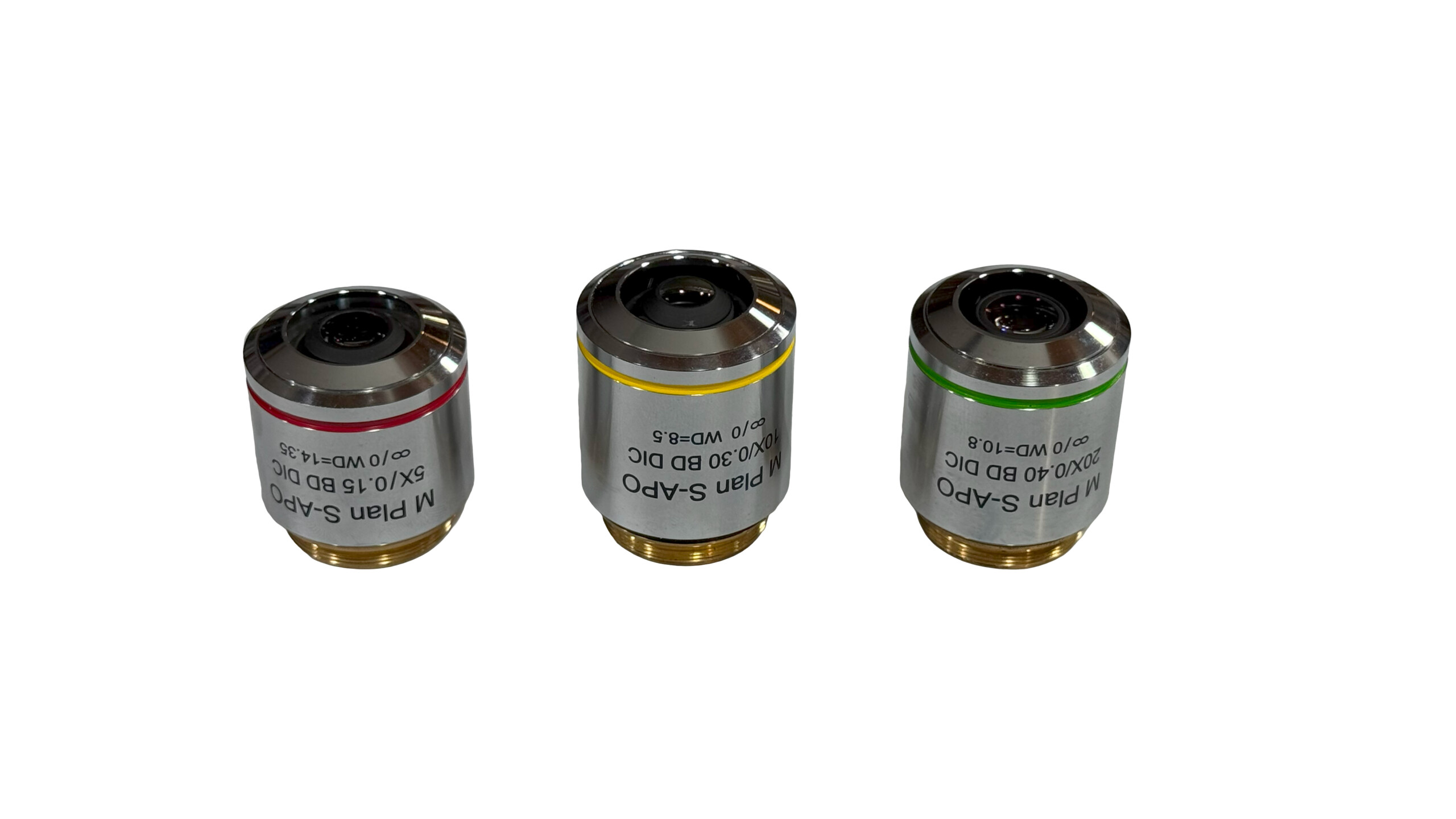
Plan Semi-Apo Lens with RMS Threads
BIA-O-5xR-BFDIC
Plan Semi-Apo
5x
0.15
20
BF, Pol, DIC
RMS
BIA-O-10xR-BFDIC
Plan Semi-Apo
10x
0.30
15
BF, Pol, DIC
RMS
BIA-O-20xR-BFDIC
Plan Semi-Apo
20x
0.40
10
BF, Pol, DIC
RMS
BIA-O-50xR-BFDIC
Plan Semi-Apo
50x
0.80
2.5
BF, Pol, DIC
RMS
Plan Semi-Apo Lens with M-26 Threads
BIA-O-2.5xM-BFDIC
Plan Semi-Apo
2.5x
0.075
6.2
BF, Pol, DIC
M26 x 0.7
BIA-O-5xM-BFDIC
Plan Semi-Apo
5x
0.15
23.5
BF, Pol, DIC
M26 x 0.7
BIA-O-10xM-BFDIC
Plan Semi-Apo
10x
0.30
22.8
BF, Pol, DIC
M26 x 0.7
BIA-O-20xM-BFDIC
Plan Semi-Apo
20x
0.40
19.2
BF, Pol, DIC
M26 x 0.7
BIA-O-50xM-BFDIC
Plan Semi-Apo
50x
0.80
11.0
BF, Pol, DIC
M26 x 0.7
Plan S-Apo Lens with M26 Threads
BIA-O-5xM-BDDIC
Plan S-Apo
5x
0.15
14.8
BF, DF, Pol, DIC
M26 x 0.7
BIA-O-10xM-BDDIC
Plan S-Apo
10x
0.30
8.5
BF, DF, Pol, DIC
M26 x 0.7
BIA-O-20xM-BDDIC
Plan S-Apo
20x
0.40
10.8
BF, DF, Pol, DIC
M26 x 0.7
BIA-O-50xM-BDDIC
Plan S-Apo
50x
0.75
3.0
BF, DF, Pol, DIC
M26 x 0.7
BIA-O-100xM-BDDIC
Plan S-Apo
100x
0.8
1.0
BF, DF, Pol, DIC
M26 x 0.7
Dimensions
Application
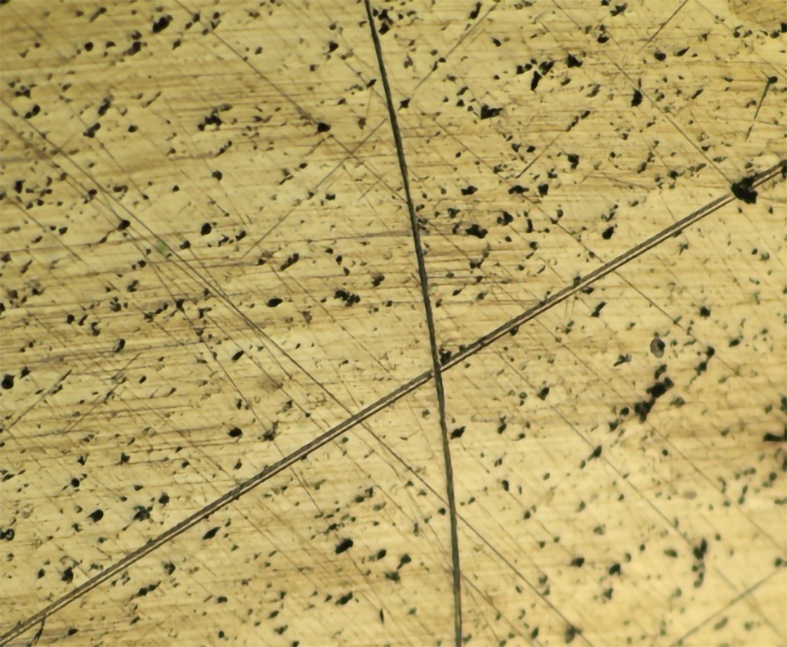
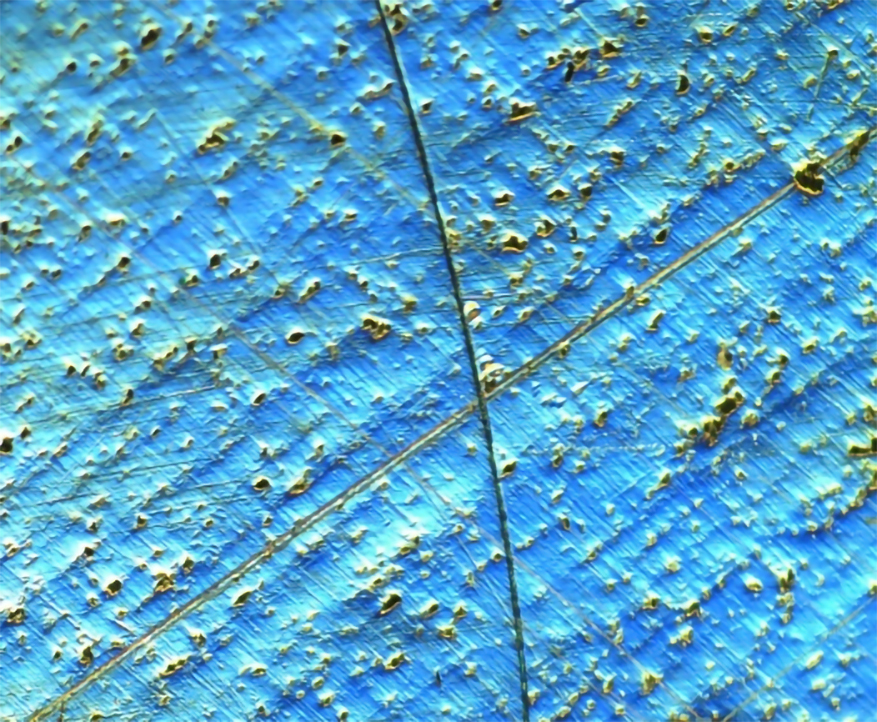
1. Conductive Particle Detection in LCD/OLED and Other Products
Detecting conductive particles in LCD circuits is crucial for ensuring optimal conductive performance. An insufficient number of conductive particles can degrade the circuit’s conductivity, potentially leading to display failures. Conversely, an excess can result in raw material wastage. Additionally, adhered conductive particles can skew particle counts, leading to underestimation and inaccurate detection results.
As illustrated in Figure 1, conductive particles are not visible using a metallographic microscope with reflected light brightfield imaging. In contrast, Figure 2 demonstrates the clear outline of these particles when using a DIC microscope system. Figure 2 depicts conductive particles on an LCD screen captured with a DIC microscope, while Figure 1 shows the same area imaged with a metallographic microscope.
This highlights the superior capability of DIC microscopy in accurately detecting and analyzing conductive particles, making it an indispensable tool for researchers and professionals in the field.
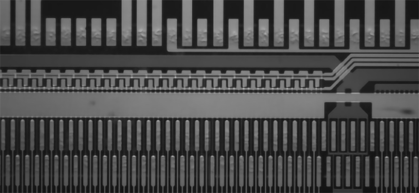
Figure 1A. Brightfield, Blue LED, Monochrome Camera
Figure 1B. Brightfield, White LED, Color Camera
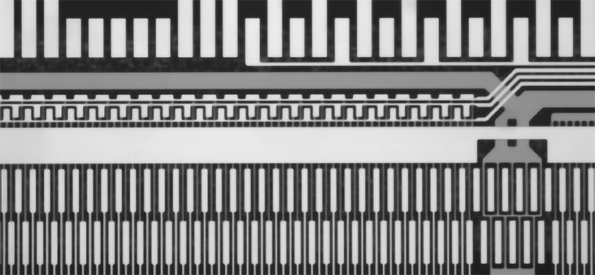
Figure 2A. DIC, Blue LED, Monochrome Camera
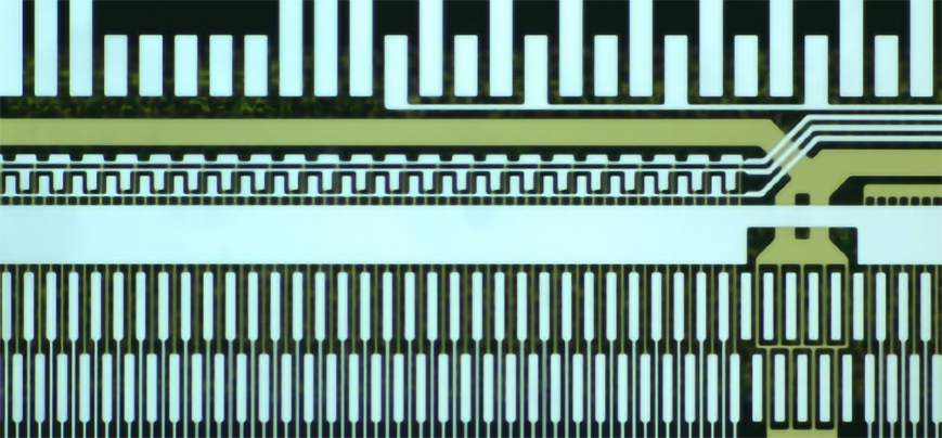
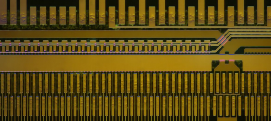
Figure 2B. DIC, White LED, Color Camera
2. Sample Surface Crack and Defect Detection
Differential Interference Contrast (DIC) microscopy stands out as a powerful tool in modern material metallographic examination, offering several advantages. It requires relatively low sample preparation and provides a pronounced relief effect in observed images.
Figure 3 illustrates this capability: the left side shows fine structures or defects that are invisible or barely visible under an ordinary metallographic microscope with incident light, while the right side reveals these details clearly using the MVM-DIC Series Differential Interference Contrast microscope system. Additionally, the MVM-DIC series system excels at revealing particles, holes, cracks, and uneven contours in the sample, ensuring more reliable material analysis.
This makes the MVM-DIC Series an indispensable instrument for researchers and professionals focused on detailed and accurate detection of surface cracks and defects.
Figure 3A. Brightfield
Figure 3B. DIC Nomarski
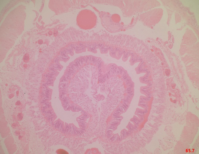
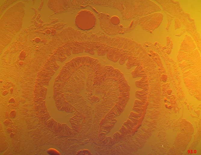
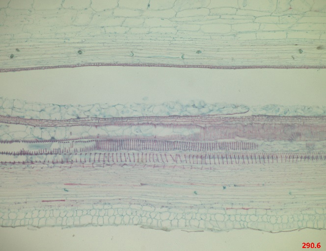
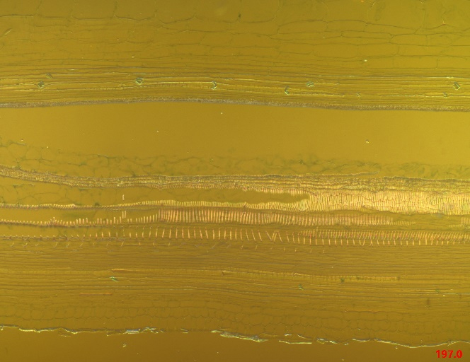
3. Microbial Cell Detection
The BVM-DIC Series Differential Interference Contrast microscope system enables non-destructive detection of living cell activity. Utilizing optical staining effects, it allows for the adjustment of images with different interference colors and the ability to change the focal length to obtain clear images at various depths. With high resolution, this system can distinctly display intracellular contours and structures. Figures 4 and 5 demonstrate the comparative results, highlighting the effectiveness of the BVM-DIC system in microbial cell detection.
This advanced system is ideal for researchers and academics focused on precise and detailed observations of living microbial cells.
Figure 4A. Earthworm, Brightfield, 50x
Figure 4B. Earthworm, DIC, 50x
Figure 5A. Cucurbit Stem, brightfield
Figure 5B. Cucurbit Stem, DIC
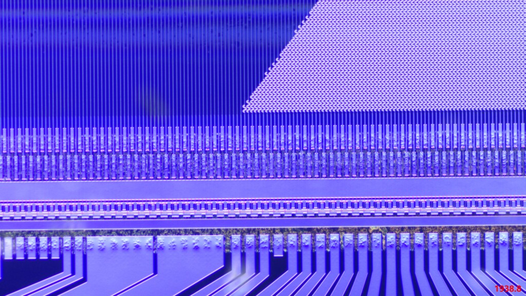
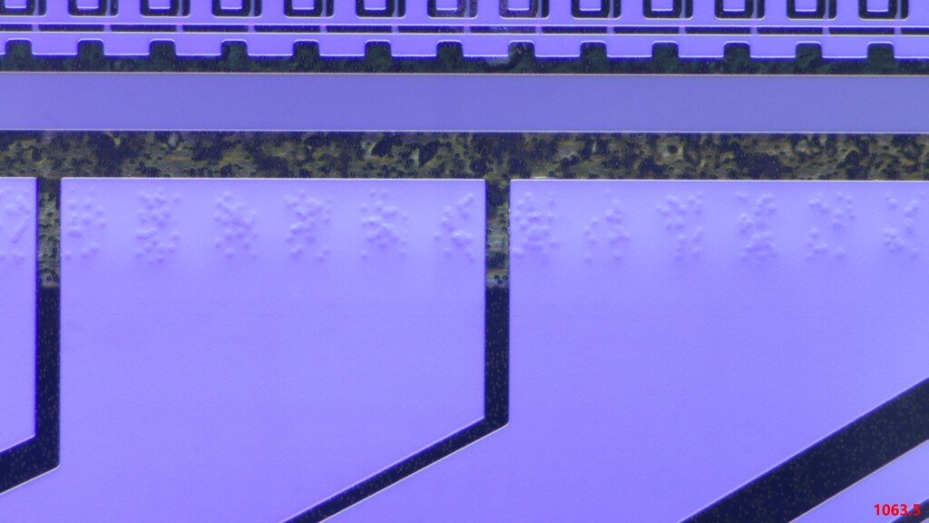
Figure 6A. Semiconductor, 10x DIC lens, a 8.3MP camera
Figure 6A. Semiconductor, 20x DIC lens, a 8.3MP camera


Specifications